The human heart consists largely of elongated muscle cells, some 100 ?m long and 15 ?min diameter.
Question:
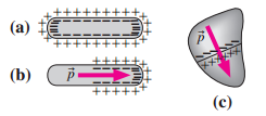
The human heart consists largely of elongated muscle cells, some 100 ?m long and 15 ?min diameter. In its resting state, a cell contains two concentric layers of charge, which confine the electric field to the cell membrane (Fig. 20.38a). When the heart contracts, a wave of depolarization sweeps through, depleting charge and giving each cell a dipole moment (Fig. 20.38b). As a result, the entire organ acts like an electric dipole, producing an external field, which is indirectly ? detected by electrocardiography. Although the direction of the heart?s dipole moment varies, Fig. 20.38c is typical. In answering the ?questions that follow, consider the heart in isolation don?t concern yourself with the effect of surrounding tissues on its electric field.

Figure 20.38 ?Heart cells (a) in the resting state and (b) partially depolarized, resulting in a dipole moment pu. (c) Typical orientation of the heart?s dipole moment vector. Cells along the line are depolarizing.
At the instant shown in Fig. 20.38c, there?s an electric field within the heart that points approximatelya. ?in the direction of the dipole moment vector p.b. ?opposite the dipole moment vector p.c. ?perpendicular to the dipole moment vector p.
Step by Step Answer:


