Question
Brain activations were measured during the presentations of negative (unpleasant) and neutral pictures in a functional magnetic resonance imaging (fMRI) study. After this initial presentation,
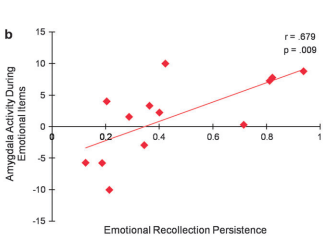
Brain activations were measured during the presentations of negative (unpleasant) and neutral pictures in a functional magnetic resonance imaging (fMRI) study. After this initial presentation, the participants were shown some of the previously shown pictures as well as new pictures. The researchers then evaluated the relationship between amygdala (a brain area important for processing emotion) activation and memory performance (how well they remembered the negative, unpleasant images). Amygdala activity scores are on the y-axis and emotional memory persistence (or how long negative emotion memories last) is on the x-axis. The graph below shows the results of the study. The correlation analysis produced: r=0.679, p=0.009.

Step by Step Solution
There are 3 Steps involved in it
Step: 1

Get Instant Access to Expert-Tailored Solutions
See step-by-step solutions with expert insights and AI powered tools for academic success
Step: 2

Step: 3

Ace Your Homework with AI
Get the answers you need in no time with our AI-driven, step-by-step assistance
Get Started


