

Antibody labeling of proteins like that used in immune-fluorescence analysis can be applied to electron microscopy, but
Question:
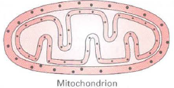
a. Using this approach, investigators have determined the subcellular localization of Tim and Tom proteins used for protein import into mitochondria. In the drawing of the results below, label the gold particles showing the localization of Tim44 and Tom40. What made you come to these conclusions?
b. Genetically engineering the N-terminus of alcohol dehydrogenase (ADH) onto a cytosolic protein alters that protein's localization. The following blot was seen when cells were transfected with an ADH-actin chimeric construct, and proteins were isolated from the cytosol and mitochondria. Antibodies against actin (43 kDa), the cytosolic protein GAPDH (37 kDa), and the mitochondrial inner membrane protein succinate dehydrogenase A (72 kDa), were used in the analysis.
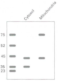
How do you explain the presence of actin in two distinct sub-cellular pools of protein? How do you explain it being the same molecular mass in both pools? If the blot were stripped and repro bed with an antibody against the N-terminus of alcohol dehydrogenase; where would you expect to find the signal?
c. Cyanide is toxic to cells because it inhibits a specific mitochondrial protein complex that is responsible for producing ATP. An experiment like that described above was repeated and the results compared to those for cells exposed to hydrogen cyanide. The following are the results from the immunoblot analysis:
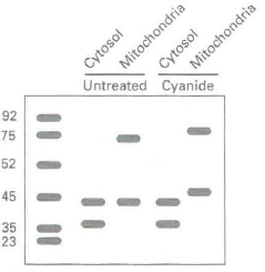
How do you explain the apparent shift in the molecular mass of actin in the mitochondrial fraction of cyanide-treated cells? Note that there is a similar shift in the mass of the succinate dehydrogenase A control. What would you expect to see on blots if cyanide-treated cells were supplemented with ATP?
Step by Step Answer:
a The smallersized gold particles are labeled Tim44 i inner and the larger particles Tom40 o outer b ...View the full answer


Molecular Cell Biology
ISBN: 978-1429234139
7th edition
Authors: Harvey Lodish, Arnold Berk, Chris A. Kaiser, Monty Krieger, Anthony Bretscher, Hidde Ploegh, Angelika Amon, Matthew P. Scott
