Answered step by step
Verified Expert Solution
Question
1 Approved Answer
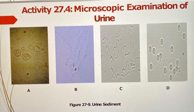
1. Observe the urine sediment images in Figure 27-9 in order to detect the presence of leukocytes, epithelial cells, mucus threads, bacteria, casts, and various
1. Observe the urine sediment images in Figure 27-9 in order to detect the presence of leukocytes, epithelial cells, mucus threads, bacteria, casts, and various crystals.
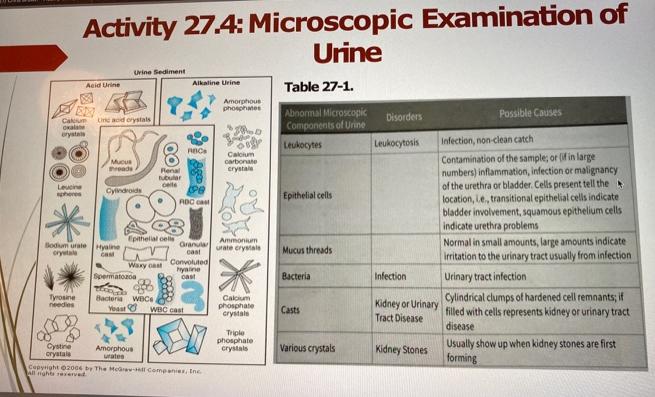
2. Table 27-1 shows abnormal components of urine, disorders, and their possible causes.
3. Identify the abnormal components in each of the urine sediment images A,B, C, and D and name the disorder associated with the presence of the components and the possible causes of the presence of the components in the urine samples.


Leucine EX Calum Line acid crystals Oxalate crystals spheres "** Activity 27.4: Microscopic Examination of Urine Acid Urine Tyrosine needles Cystine crystals Mucus hreads Sodium urate Hyaline crystal cast Urine Sediment Cylindroids Epithelial cells Waxy cast Spermatozoa Renal tubular celle Bacteria WDC Yosar Amorphous urates Alkaline Urine ROC PB ADC cast Granular cast WBC cast Convoluted hyaline cast Copyright 2006 by The McGraw-Hill Companies, Inc. All rights reserved. Amorphous phosphates D OB Calcium carbonate crystal Ammonium urate crystals Calcium phosphate crystals Triple phosphate crystals Table 27-1. Abnormal Microscopic Components of Urine Leukocytes Epithelial cells Mucus threads Bacteria Casts Various crystals Disorders Leukocytosis Infection Possible Causes Normal in small amounts, large amounts indicate irritation to the urinary tract usually from infection Urinary tract infection Cylindrical clumps of hardened cell remnants; if Kidney or Urinary filled with cells represents kidney or urinary tract Tract Disease disease Usually show up when kidney stones are first forming Kidney Stones Infection, non-clean catch Contamination of the sample; or (if in large numbers) inflammation, infection or malignancy. of the urethra or bladder. Cells present tell the location, L.e., transitional epithelial cells indicate bladder involvement, squamous epithelium cells indicate urethra problems
Step by Step Solution
★★★★★
3.45 Rating (152 Votes )
There are 3 Steps involved in it
Step: 1
Microscopic examination of Urine A Abnormal components i...
Get Instant Access to Expert-Tailored Solutions
See step-by-step solutions with expert insights and AI powered tools for academic success
Step: 2

Step: 3

Ace Your Homework with AI
Get the answers you need in no time with our AI-driven, step-by-step assistance
Get Started


