

Answered step by step
Verified Expert Solution
Question
1 Approved Answer
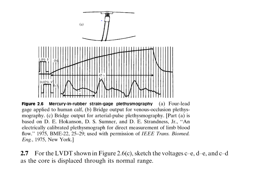
1.0% (b 45 s 0.02% V Figure 2.6 Mercury-in-rubber strain-gage plethysmography (a) Four-lead gage applied to human calf, (b) Bridge output for venous-occlusion plethys- mography.
Step by Step Solution
There are 3 Steps involved in it
Step: 1

Get Instant Access to Expert-Tailored Solutions
See step-by-step solutions with expert insights and AI powered tools for academic success
Step: 2
Step: 3
Ace Your Homework with AI
Get the answers you need in no time with our AI-driven, step-by-step assistance
Get Started
Database Design And Relational Theory Normal Forms And All That Jazz
Authors: Chris Date
1st Edition
1449328016, 978-1449328016