Answered step by step
Verified Expert Solution
Question
1 Approved Answer
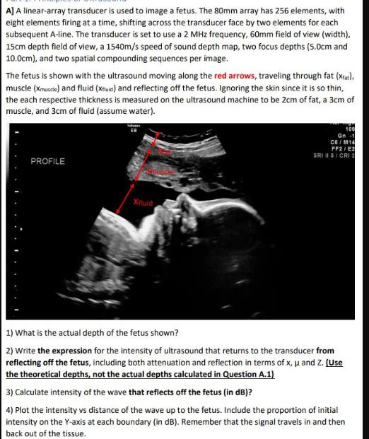
A] A linear-array transducer is used to image a fetus. The 80mm array has 256 elements, with eight elements firing at a time, shifting

A] A linear-array transducer is used to image a fetus. The 80mm array has 256 elements, with eight elements firing at a time, shifting across the transducer face by two elements for each subsequent A-line. The transducer is set to use a 2 MHz frequency, 60mm field of view (width), 15cm depth field of view, a 1540m/s speed of sound depth map, two focus depths (5.0cm and 10.0cm), and two spatial compounding sequences per image. The fetus is shown with the ultrasound moving along the red arrows, traveling through fat (xat), muscle (Xmuscle) and fluid (Xuid) and reflecting off the fetus. Ignoring the skin since it is so thin, the each respective thickness is measured on the ultrasound machine to be 2cm of fat, a 3cm of muscle, and 3cm of fluid (assume water). PROFILE Ya EB Sydsche Xfluid 100 Gn 1 C6/M14 FF2/E2 SRI II S/CRI 2 1) What is the actual depth of the fetus shown? 2) Write the expression for the intensity of ultrasound that returns to the transducer from reflecting off the fetus, including both attenuation and reflection in terms of x, and Z. (Use the theoretical depths, not the actual depths calculated in Question A.1) 3) Calculate intensity of the wave that reflects off the fetus (in dB)? 4) Plot the intensity vs distance of the wave up to the fetus. Include the proportion of initial intensity on the Y-axis at each boundary (in dB). Remember that the signal travels in and then back out of the tissue.
Step by Step Solution
There are 3 Steps involved in it
Step: 1

Get Instant Access to Expert-Tailored Solutions
See step-by-step solutions with expert insights and AI powered tools for academic success
Step: 2

Step: 3

Ace Your Homework with AI
Get the answers you need in no time with our AI-driven, step-by-step assistance
Get Started


