I would like you to point out all the problems with this essay which include factual errors, and issues with referencing ets. make a list
I would like you to point out all the problems with this essay which include factual errors, and issues with referencing ets. make a list of all those things and provide Re1ferences on where you got those resources from, Ideally pubmed.
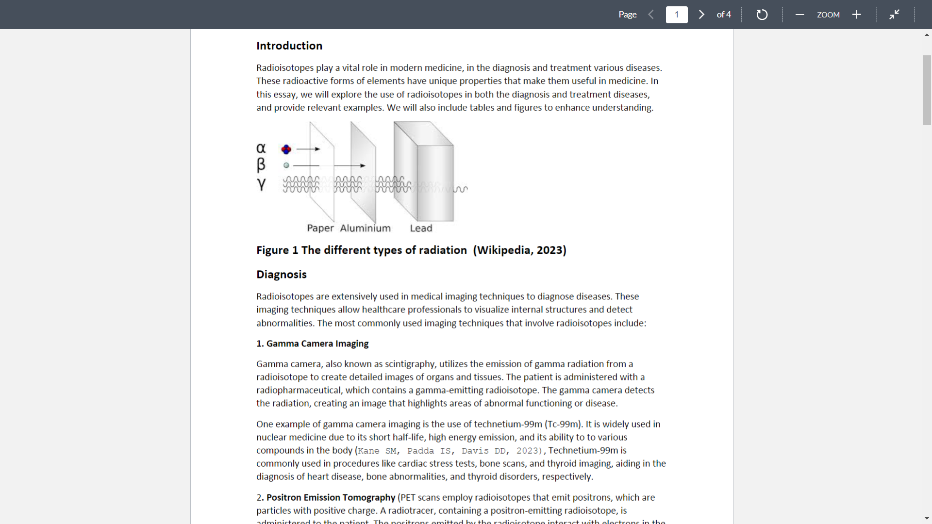
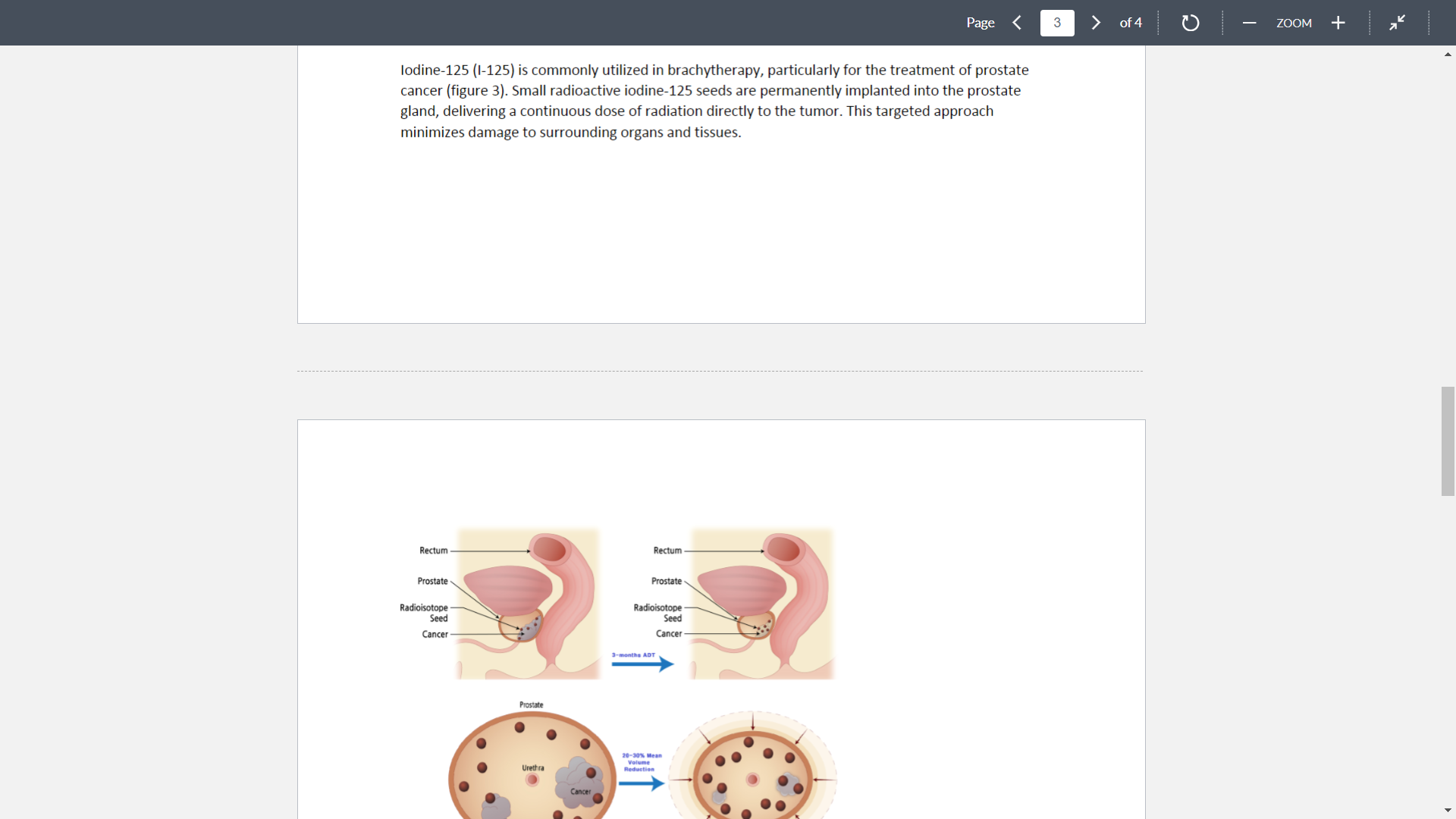
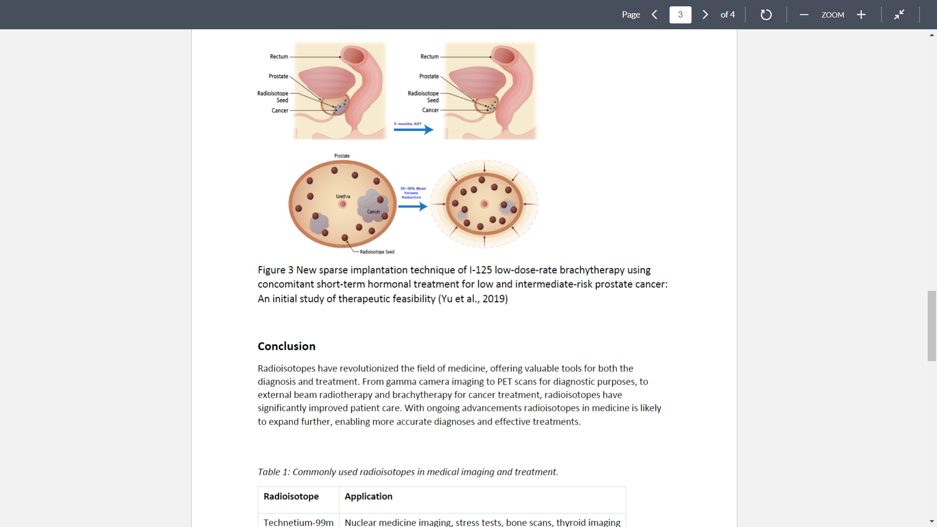
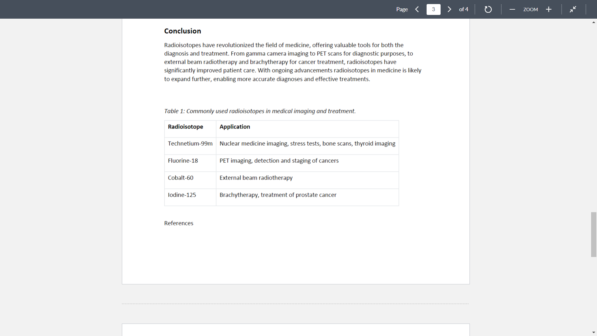
> of4 D ZOOM + Page Introduction Radioisotopes play a vital role in modern medicine, in the diagnosis and treatment various diseases. These radioactive forms of elements have unique properties that make them useful in medicine. In this essay, we will explore the use of radioisotopes in both the diagnosis and treatment diseases, and provide relevant examples. We will also include tables and figures to enhance understanding. Paper Aluminium Lead Figure 1 The different types of radiation (Wikipedia. 2023) Diagnosis Radioisotopes are extensively used in medical imaging techniques to diagnose diseases. These imaging techniques allow healthcare professionals to visualize internal structures and detect abnormalities. The most commonly used imaging techniques that involve radioisotopes include: 1. Gamma Camera Imaging Gamma camera, also known as scintigraphy, utilizes the emission of gamma radiation from a radioisotope to create detailed images of organs and tissues, The patient is administered with a radiopharmaceutical, which contains a gamma-emitting radioisotope. The gamma camera detects the radiation, creating an image that highlights areas of abnormal functioning or disease. One example of gamma camera imaging is the use of technetium-99m (Tc-99m). It is widely used in nuclear medicine due to its short half-life, high energy emission, and its ability to to various compounds in the body (Kane SM, Patina IS, Dans :D, 2323),Technetium-99m is commonly used in procedures like cardiac stress tests, bone scans, and thyroid imaging, aiding in the diagnosis of heart disease, bone abnormalities, and thyroid disorders, respectively. 2. Positron Emission Tomography (PET scans employ radioisotopes that emit positrons, which are particles with positive charge. A radiotracer, containing a positroneemitting radioisotope, is summhpnma on \"M "sun\" rim "mam" \"mm\": L.\" um \".4: \"sh...\" znpmvs mm. \"arena" :n u... 2. Positron Emission Tomography (PET scans employ radioisotopes that emit positrons, which are particles with positive charge. A radiotracer, containing a positron-emitting radioisotope, is administered to the patient. The positrons emitted by the radioisotope interact with electrons in the body, resulting in the emission of gamma rays. The PET scanner detects these gamma rays and constructs a threeedimensional image, offering insights into the metabolic and biochemical processes within the body. Fluorine-18 (F-18) is commonly used in PET imaging. For example, fluorodeoxyglucose (FDGJ, a glucose analog labeled with F718, is used to visualize glucose metabolism in the body, including for the diagnosis of autoimmune encephalitis (Bordonne et al, 2021). PET scans using FDG can help detect and stage various cancers, such as lung, colon, and lymph. Treatment Radioisotopes also have therapeutic applications in the treatment of diseases, particularly cancer (Voon et al. 2017). Radioactive substances can be targeted to certain areas of the body a concentrated dose of radiation, specifically to cancer cells. The two main techniques involving the use of radioisotopes for treatment are: 1. External Beam Radiotherapy External beam radiotherapy involves the use of a machine that generates higheenergy radiation, such as Xerays or electrons. These beams are directed towards the tumor from outside the body, with the aim of destroving cancer cells. Radioactive sources. such as cobalte (@760) or linear Page of4 D ZOOM + 1. External Beam Radiotherapy External beam radiotherapy involves the use of a machine that generates high-energy radiation, such as Xerays or electrons. These beams are directed towards the tumor from outside the body, with the aim of destroying cancer cells. Radioactive sources, such as cobalteGU (C0760) or linear accelerators, are used to generate the radiation. This is commonly used to treat bone metastases (Ejima et al, 2017 New Paradigms of Radiotherapy for Bone Metastasis) One example of external beam radiotherapy is intensity-modulated radiation therapy (IMRT). This technique utilizes multiple radiation beams of different intensities to precisely target the tumor while minimizing damage to surrounding healthy tissues. By adjusting the intensity of each beam, IMRT allows for high doses of radiation to the tumor while sparing critical structures nearby. 2. Internal Radiotherapy (Brachytherapy) In internal radiotherapy, also known as brachytherapv, a radioactive source is placed near or inside the tumor (Mayer et al. 2023). This allows for direct application of radiation to the tumor site, minimizing exposure to healthy tissues. The radioactive material can be delivered via seeds, wires, or catheters, depending on the specific treatment plan (Figure 2)' Figure 2 Diagram showing hrachytherapy administration to the lung (CIRSE) Brachytherapy (example in the lung) Catheter Radioactive source Tumour Lung Iodine7125 (HZS) is commonly uti MMMMMM no... m cm-" Moan: ed in brachvtherapy, particularly for the treatment of prostate "4:,\" aw: \"ma, -m nmmvmnnu \"Masai\": :ntn u\" \"we.\" Page 3 of 4 - ZOOM + lodine-125 (1-125) is commonly utilized in brachytherapy, particularly for the treatment of prostate cancer (figure 3). Small radioactive iodine-125 seeds are permanently implanted into the prostate gland, delivering a continuous dose of radiation directly to the tumor. This targeted approach minimizes damage to surrounding organs and tissues. Rectum Rectum Prostate Prostate Radioisotope Radioisotope Seed Seed Cancer Cancer -months ADT Prostate 20-30%% Mea Urethra Reduction CancePage
Step by Step Solution
There are 3 Steps involved in it
Step: 1
Get Instant Access to Expert-Tailored Solutions
See step-by-step solutions with expert insights and AI powered tools for academic success
Step: 2
Step: 3
Ace Your Homework with AI
Get the answers you need in no time with our AI-driven, step-by-step assistance