Answered step by step
Verified Expert Solution
Question
1 Approved Answer
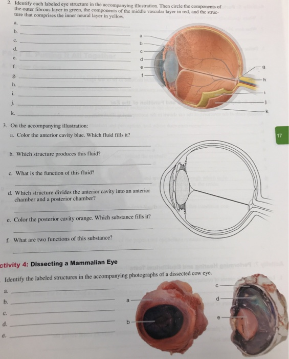
2. Identify each labeled eye structure in the accompanying illustration. Then circle the components of the outer fibrous layer in green, the components of

2. Identify each labeled eye structure in the accompanying illustration. Then circle the components of the outer fibrous layer in green, the components of the middle vascular layer in red, and the struc- ture that comprises the inner neural layer in yellow. a. b. C. a. e. d. b. e. f. 3. On the accompanying illustration: a. Color the anterior cavity blue. Which fluid fills it? C. d. g. h. i. j. k. b. Which structure produces this fluid? f. What are two functions of this substance? c. What is the function of this fluid? d. Which structure divides the anterior cavity into an anterior chamber and a posterior chamber? e. Color the posterior cavity orange. Which substance fills it? a b activity 4: Dissecting a Mammalian Eye Identify the labeled structures in the accompanying photographs of a dissected cow eye. C d e d 17
Step by Step Solution
★★★★★
3.34 Rating (145 Votes )
There are 3 Steps involved in it
Step: 1
Question 2 A CILIARY MUSCLE MIDDLE VASCULAR LAYER B SUSPENSORY LIGAMENT MIDDLE VASCULAR LAYER C IRIS ...
Get Instant Access to Expert-Tailored Solutions
See step-by-step solutions with expert insights and AI powered tools for academic success
Step: 2

Step: 3

Ace Your Homework with AI
Get the answers you need in no time with our AI-driven, step-by-step assistance
Get Started


