Answered step by step
Verified Expert Solution
Question
1 Approved Answer
Part 1: Gram Stain Procedure Procedure 1. Examine the finished slides under a microscope using the 10X, 40X, and the oil immersion (100X) lenses
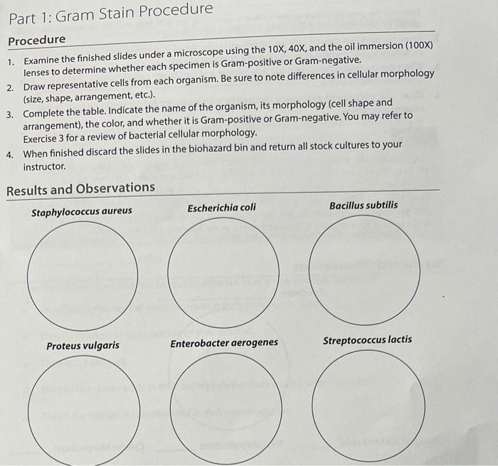
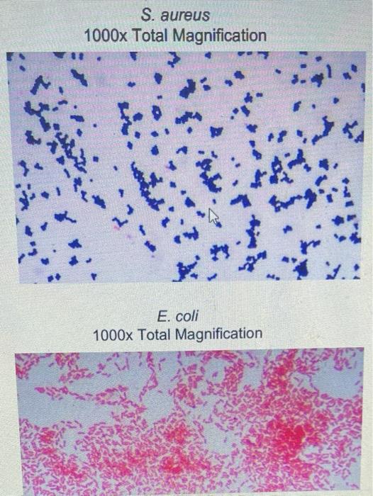
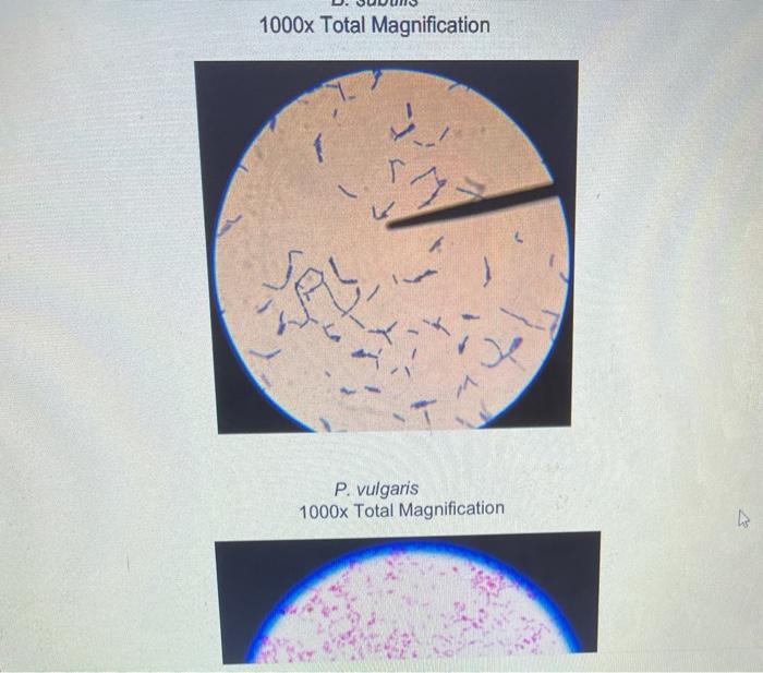
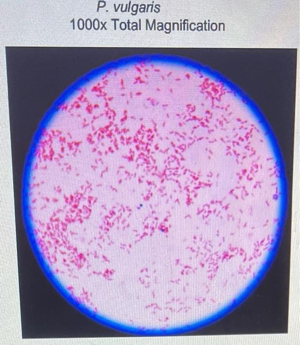
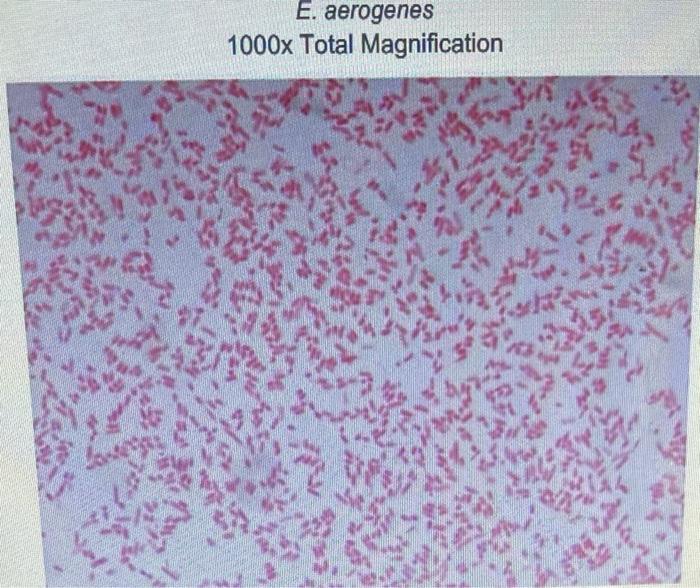
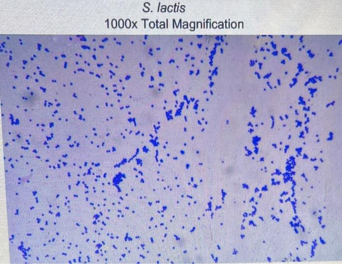
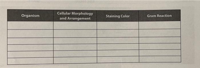
Part 1: Gram Stain Procedure Procedure 1. Examine the finished slides under a microscope using the 10X, 40X, and the oil immersion (100X) lenses to determine whether each specimen is Gram-positive or Gram-negative. 2. Draw representative cells from each organism. Be sure to note differences in cellular morphology (size, shape, arrangement, etc.). 3. Complete the table. Indicate the name of the organism, its morphology (cell shape and arrangement), the color, and whether it is Gram-positive or Gram-negative. You may refer to Exercise 3 for a review of bacterial cellular morphology. 4. When finished discard the slides in the biohazard bin and return all stock cultures to your instructor. Results and Observations Staphylococcus aureus Proteus vulgaris Escherichia coli Enterobacter aerogenes Bacillus subtilis Streptococcus lactis S. aureus 1000x Total Magnification 77 KI E. coli 1000x Total Magnification Mo 3.1 1000x Total Magnification T P. vulgaris 1000x Total Magnification P. vulgaris 1000x Total Magnification E. aerogenes 1000x Total Magnification 946 S. lactis 1000x Total Magnification Organism Cellular Morphology and Arrangement Staining Color Gram Reaction
Step by Step Solution
★★★★★
3.33 Rating (147 Votes )
There are 3 Steps involved in it
Step: 1
1 S aureus Cellular morphology and arrangement cocci cluster tetrads arrang...
Get Instant Access to Expert-Tailored Solutions
See step-by-step solutions with expert insights and AI powered tools for academic success
Step: 2

Step: 3

Ace Your Homework with AI
Get the answers you need in no time with our AI-driven, step-by-step assistance
Get Started


