Answered step by step
Verified Expert Solution
Question
1 Approved Answer
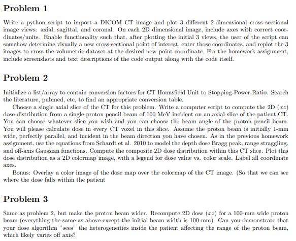
Problem 1 Write a python script to import a DICOM CT image and plot 3 different 2 - dimensional cross sectional image views: axial, sagittal,
Problem
Write a python script to import a DICOM CT image and plot different dimensional cross sectional
image views: axial, sagittal, and coronal. On each D dimensional image, include axes with correct coor
dinatesunits Enable functionality such that, after plotting the initial views, the user of the script can
somehow determine visually a new crosssectional point of interest, enter those coordinates, and replot the
images to cross the volumetric dataset at the desired new point coordinate. For the homework assignment,
include screenshots and text descriptions of the code output along with the code itself.
Problem
Initialize a listarray to contain conversion factors for CT Hounsfield Unit to StoppingPowerRatio. Search
the literature, pubmed, etc, to find an appropriate conversion table.
Choose a single axial slice of the CT for this problem. Write a computer script to compute the D
dose distribution from a single proton pencil beam of MeV incident on an axial slice of the patient CT
You can choose whatever slice you wish and you can choose the beam angle of the proton pencil beam.
You will please calculate dose in every CT voxel in this slice. Assume the proton beam is initially
wide, perfectly parallel, and incident in the beam direction you have chosen. As in the previous homework
assignment, use the equations from Schardt et al to model the depth dose Bragg peak, range straggling,
and offaxis Gaussian functions. Compute the composite D dose distribution within this CT slice. Plot this
dose distribution as a D colormap image, with a legend for dose value vs color scale. Label all coordinate
axes.
Bonus: Overlay a color image of the dose map over the colormap of the CT image. So that we can see
where the dose falls within the patient
Problem
Same as problem but make the proton beam wider. Recompute D dose for a mm wide proton
beam everything the same as above except the initial beam width is Can you demonstrate that
your dose algorithm "sees" the heterogeneities inside the patient affecting the range of the proton beam,
which likely varies off axis?

Step by Step Solution
There are 3 Steps involved in it
Step: 1

Get Instant Access to Expert-Tailored Solutions
See step-by-step solutions with expert insights and AI powered tools for academic success
Step: 2

Step: 3

Ace Your Homework with AI
Get the answers you need in no time with our AI-driven, step-by-step assistance
Get Started


