Answered step by step
Verified Expert Solution
Question
1 Approved Answer
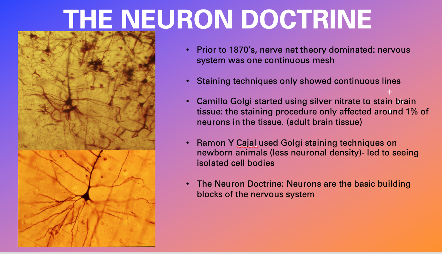
THE NEURON DOCTRINE Prior to 1870's, nerve net theory dominated: nervous system was one continuous mesh Staining techniques only showed continuous lines + Camillo


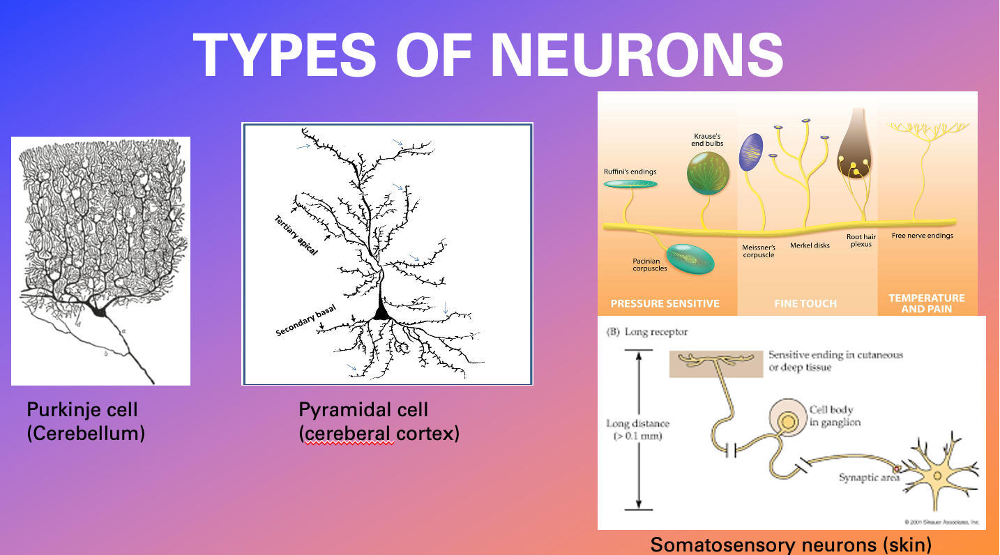
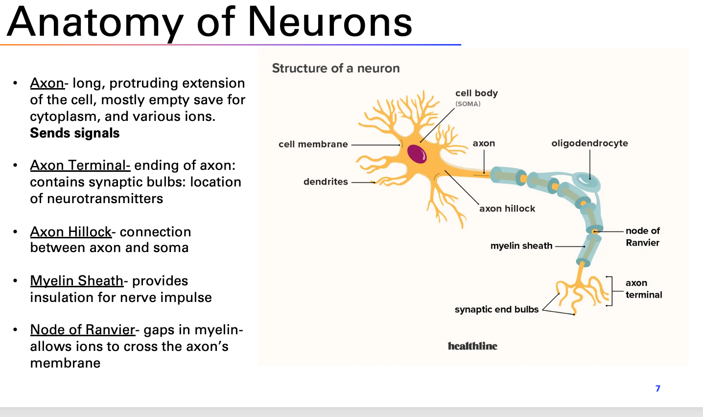
THE NEURON DOCTRINE Prior to 1870's, nerve net theory dominated: nervous system was one continuous mesh Staining techniques only showed continuous lines + Camillo Golgi started using silver nitrate to stain brain tissue: the staining procedure only affected around 1% of neurons in the tissue. (adult brain tissue) Ramon Y Cajal used Golgi staining techniques on newborn animals (less neuronal density)- led to seeing isolated cell bodies The Neuron Doctrine: Neurons are the basic building blocks of the nervous system TYPES OF NEURONS Tertiary apical Purkinje cell (Cerebellum) Secondary basal Pyramidal cell (cereberal cortex) Ruffini's endings Pacinian corpuscles Krause's end bulbs Meissner's corpuscle Merkel disks PRESSURE SENSITIVE FINE TOUCH Root hair plexus Free nerve endings TEMPERATURE AND PAIN (B) Long receptor Sensitive ending in cutaneous or deep tissue Long distance (>0.1 mm) Cell body in ganglion Synaptic area 200 sociones, ne Somatosensory neurons (skin) Anatomy of Neurons Axon- long, protruding extension of the cell, mostly empty save for cytoplasm, and various ions. Sends signals Axon Terminal- ending of axon: contains synaptic bulbs: location of neurotransmitters Axon Hillock- connection between axon and soma Structure of a neuron cell membrane dendrites Myelin Sheath- provides insulation for nerve impulse cell body (SOMA) axon oligodendrocyte axon hillock myelin sheath node of Ranvier Node of Ranvier- gaps in myelin- allows ions to cross the axon's membrane synaptic end bulbs healthline axon terminal 7 NEURONAL SIGNALING Vagus Nerve Heart 2 Two initial schools of thought Sparks- neuronal signaling was electrical: evidenced by microelectrodes to observe neurons signaling "Soups"- Neurons communicated with chemicals Otto Loewi's frog heart experiment (came to him in a dream) Stimulated one frog heart's vagus nerve with electricity, increased beating of the heart Transferred some of the solution Heart "A" was into a dish Heart "B" was in- heart "B" started beating faster as well! NEURONAL SIGNALING Things to note: the action potential, is all or nothing: if the + cell depolarizes enough to trigger the action potential, the cell transmits it signal. Graded potentials (not full potentials) occur in the cell receiving the transition. Therefore-fine tuning (modulation) of cell signaling requires both excitatory and inhibitory synapses 2000 UTHSCH MicroNetwork Motifs A. Feedforward excitation D. Lateral inhibition B. Feedforward inhibition Excitation Inhibition E. Feedback/Recurrent inhibition F. Feedback/Recurrent excitation E1 F1 F2 C. Convergence/divergence E2 Figure 6 NEURONS AND COMPLEX STIMULI Gross et al (1972) Monkeys were shown various images while single neurons in their temporal lobes were monitored The study found that rather than simple lines, neurons in the temporal lobe responded to more complex stimuli (ex: a hand) Shows Hierarchical Processing- visual stimuli progresses along a pathway, first being processed simply, then more complex the further along the path the information travels. Receptive fields size Features IT faces LGN edges V4 V1 and lines V2 objects shapes edges shapes V1 faces and lines and objects visual field Anatomy of the Brain Temporal lobe Managing emotions, processing information from senses (heavily responsible for hearing), storing/ retrieving memories and understanding language Works closely with limbic system temporal (hippocampus and amygdala) to create associations between memories and emotions pole What? TEMPORAL LOBE SUBSTRUCTURES -limbic system region Wernicke's Area auditory cortex superior temporal gyrus middle temporal gyrus - inferior temporal gyrus 27 What happens when areas are damaged? (Temporal lobe) Difficulties in understanding spoken language (receptive aphasia- Wernicke's aphasia) Difficulties in selective attention to what we hear Difficulties in identification and categorization of objects, Difficulty learning and retaining new information Temporal lobe epilepsy can create visions, intense emotions and beliefs. Historical figures who may have had TLE: Joan of Arc, Tolstoy Localized Representation (by measurement) By using fMRI, we have been able to see what specific areas of the brain react to certain tasks or respond to different stimuli Fusiform face area (FFA)- activates when looking at faces Para hippocampal place area (PPA)- activates in response to seeing indoor and outdoor scenes Extrastriate body area (EBA)- activated when sing pictures of bodies and body parts
Step by Step Solution
There are 3 Steps involved in it
Step: 1

Get Instant Access to Expert-Tailored Solutions
See step-by-step solutions with expert insights and AI powered tools for academic success
Step: 2

Step: 3

Ace Your Homework with AI
Get the answers you need in no time with our AI-driven, step-by-step assistance
Get Started


