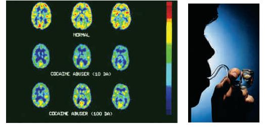
In PET scans, red areas are brain regions that are most active, while blue, yellow, and green
Question:
In PET scans, red areas are brain regions that are most active, while blue, yellow, and green areas are least active. Figure 13.28 shows PET scans of normal brain activity (left) and of the brain of a person while using cocaine (right). The frontal lobes of the brain hemispheres are toward the top of the scans. Their neurons play major roles in reasoning and other intellectual functions. Looking at these scan images, how do you suppose cocaine may affect mental functioning?
Figure 13.28.

Fantastic news! We've Found the answer you've been seeking!
Step by Step Answer:
Related Book For 

Question Posted:


